


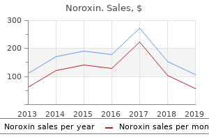
"400mg noroxin, antibiotic resistance ontology".
By: B. Milten, M.A., M.D., M.P.H.
Clinical Director, Osteopathic Medical College of Wisconsin
This results in the development of antibodies which enter the foetal circulation causing haemolysis and anaemia antimicrobial finish discount noroxin 400 mg overnight delivery. When severe this may result in soft tissue oedema 7 bacteria cheap noroxin 400mg free shipping, pleural effusions antibiotics on birth control buy noroxin without prescription, ascites and pericardial effusions antibiotic for acne effective 400 mg noroxin. C Clinical Presentation Hyaline Membrane Disease: Clinically these infants are usually symptomatic within minutes of birth, with grunting, nasal flaring, intercostals retraction, tachypnoea 314 Chest, Neonatal and cyanosis. Prenatal steroid administration to mothers during the 2 days prior to delivery is safe and significantly reduces the incidence of the disease in premature infants. It promotes endogenous surfactant production and lung maturation in addition to inducing anti-oxidant enzymes. A similar response can occur when maternal steroid production is increased because of stress caused by prepartum maternal infection, toxaemia or other forms of prepartum stress. Transient Tachypnoea of the Newborn: Mild to moderate respiratory distress without cyanosis is typically present at birth or in the first couple of hours in this condition. Amniotic Fluid Aspiration: Tachypnoea is the most common clinical finding and its severity varies with the degree of aspiration. Meconium Aspiration Syndrome: Clinically these infants demonstrate pallor, cyanosis, apnoea, grunting and intercostal retraction. Respiratory and metabolic acidosis may develop due to hypoxaemia and hypercarbia secondary to ventilation-perfusion mismatch. Neonatal Pneumonia: the clinical signs and symptoms in neonatal pneumonia are frequently non-specific. The infant maybe listless, have pallor, apnoea, tachypnoea, tachycardia, bradycardia or feeding intolerance. These infants usually present after 48 h of birth and have a less fulminant course. Those infants who present in the first 48 h of life tend to have a more severe clinical picture of hypotension, shock, disseminated intravascular coagulation and multiorgan failure. Spontaneous Pneumothorax: Pneumothorax causes varying degrees of respiratory distress. Pleural Effusions: this condition is frequently diagnosed by antenatal ultrasonography and if large may be treated antenatally by thoracocentesis and/or thoracoamniotic shunt. After birth if the effusion is large the infants present with respiratory distress. Imaging the chest radiograph is the most useful imaging modality in the investigation of the various medical conditions which cause respiratory distress in the newborn period. It is also very important in determining the position of various tubes which are introduced during therapy, together with their complications. The most common of these is a misplaced endotracheal tube which ideally should be positioned above the level of the carina and the origin of the right upper lobe bronchus. Endotracheal and nasogastric tubes may perforate the trachea or oesophagus and cause a pneumothorax or pneumomediastinum. The position of pleural chest tubes to drain pneumothoraces and pleural effusions can also be assessed on the chest radiograph. Misplacement may result in diaphragmatic paralysis as a result of phrenic nerve damage. Positioning of intravenous and intra-arterial lines in the heart is not ideal as they may predispose the infant to thromboembolism. The venous catheters are best positioned in either the superior or inferior vena cava. Perforation of the superior vena cava resulting in hydrothorax and haemothorax has been reported. Umbilical arterial line tips are best located in the aorta proximal to the origins of the main arterial branches in order to avoid thrombosis and ischaemia to the dependant organs. Prior to commencement of assisted ventilation, typically the radiographic findings are those of underaeration of the lungs with fine granular opacification and air bronchograms which are diffuse and symmetrical.
The patient has pseudoexfoliation in the right eye and nuclear sclerotic cataracts in both eyes antimicrobial fabric noroxin 400 mg generic, right greater than left antibiotic resistance nice order noroxin 400 mg overnight delivery. I counseled the patient about the treatment of this and offered her radiation therapy along with the proper dose of steroids of 100 mg a day for a week and then taper down from the 100 mg virus hitting kids buy discount noroxin 400 mg on line. Zapata for consultation radiation therapy antibiotic resistance of helicobacter pylori in u.s. veterans discount noroxin uk, and then I think we would see her weekly as we get this problem under control. I did not feel she needed any topical medication, and she definitely does not need surgery now since there is no proptosis at this time. I counseled her about the side effects of the high dose of steroids and how to take them. The medical necessity of the services and supplies is reported with diagnostic codes. Because the services provided by hospitals are broader than those provided by physicians, hospitals are reimbursed differently than physicians. The hospital is paid for its "hospitality," ensuring the patient is housed, fed, and nurtured back to health until the patient is discharged. For example, a young woman admitted to the hospital for a normal, uncomplicated birth usually requires much less nursing care and recovery time than an elderly patient admitted for multiple organ failure in sepsis. For hospitals, the diagnosis is the key in determining the resources required by a patient. The instructions and conventions of the classification take precedence over guidelines. A joint effort between the healthcare provider and the coder is essential to achieve complete and accurate documentation, code assignment, and reporting of diagnoses and procedures. These guidelines have been developed to assist both the healthcare provider and the coder in identifying those diagnoses that are to be reported. The importance of consistent, complete documentation in the medical record cannot be overemphasized. The entire record should be reviewed to determine the specific reason for the encounter and the conditions treated. Sequencing of diagnostic codes is more crucial to reimbursement for hospitals, and the rules for diagnostic coding also differ. For example, a patient is admitted to the hospital experiencing severe chest pain and sweating, to rule out myocardial infarction. The patient requires 36 hours in the hospital to undergo intense diagnostics to determine the cause of the chest pain. During the hospitalization, resources expended on this patient are identical to resources for a patient with a myocardial infarct. Many hospitalizations are medical in nature (uncontrolled diabetes, pneumonia) while others are surgical in nature (mastectomy, total hip replacement). Procedure codes need to be sequenced properly with the principal procedure as the first-listed procedure. The principal procedure is one that is performed for definitive treatment rather than for diagnostic or exploratory purposes, or one necessary for a complication. If two procedures appear to meet this definition, then the one most closely related to the principal diagnosis should be assigned as the principal procedure. A procedure is considered to be significant if it: Is surgical in nature Carries a procedural risk Carries an anesthetic risk Requires specialized training to perform For a procedure to be significant it does not have to be performed in the operating room. Without such documentation the application of all coding guidelines is a difficult, if not impossible, task. In an outpatient setting, the term first-listed condition is used in lieu of the term principal diagnosis and is used to indicate the main reason for the visit. Additional diagnoses may be necessary in order to substantiate adjunct services (such as laboratory and radiology). There are specific guidelines that assist with the selection of the principal diagnosis. Chest pain is not coded, because it is a symptom of the definitive diagnosis of acute myocardial infarction. You must always refer to the Tabular; otherwise you will miss important notes, such as "additional character(s) required.
Noroxin 400 mg without a prescription. Textile Waste Recycling Using a Biological Method.
Prevention of this iatrogenic condition is important to avoid the substantial morbidity and even mortality that can sometimes be associated with contrast medium-induced nephrotoxicity antibiotic prophylaxis for colonoscopy buy noroxin without prescription. Even a small decrease in renal function may greatly exacerbate the morbidity and mortality caused by coexisting conditions such as acquired sepsis antibiotic yellow tongue cheap 400 mg noroxin otc, bleeding bacteria escherichia coli buy 400mg noroxin overnight delivery, coma antibiotics and drinking purchase 400 mg noroxin with amex, and respiratory failure, which are more frequent in patients with acute renal failure. Therefore, several measures have been tested to reduce the frequency of contrast medium-induced nephropathy. No measure has yet resulted in avoidance of its occurrence in all patients, but adequate hydration reduces the incidence. Synonyms Radiocontrast media; Radiographic contrast media Definition Contrast media are drugs that enhance the differences seen on the images between the body tissues. The ideal contrast agent should be totally inert, causing no interactions with the organism at any level. They can be divided into water-soluble iodinated contrast media and non-water-soluble barium agents. The water-soluble iodinated contrast media can be further subdivided according to the number of benzenerings: monomers (one ring), dimers (two rings). Thus iodinated contrast agents can be divided into four classes: (i) Highosmolar ionic monomeric agents, (ii) Low-osmolar nonionic monomeric agents, (iii) Low-osmolar ionic dimeric agents, and (iv) Iso-osmolar non-ionic dimeric agents. Interactions Several of the in vitro studies on the hematologic effects of contrast media have not been confirmed by in vivo and clinical studies. They can also be administered directly into the gastrointestinal tract and urinary tract, or into body cavities to opacify fistulas for example. Barium sulphate preparations used to visualize the gastrointestinal tract consist of a suspension of insoluble barium sulphate particles which are not absorbed from the gut. Contraindications Iodinated Contrast Media Absolute: All contrast media: manifest hyperthyroidism Ionic contrast media: subarachnoidal injection. Relative: Reduced renal function, asthma, history of allergy, previous reaction to a iodinated contrast medium Pregnancy/Lactation If the examination has been found to be indicated based on a thoroughly analysis of the history, symptoms and signs, one should go ahead and use the contrast media whenever it is necessary in order to confirm the presence or absence of a lesion. In early pregnancy, mutagenic and teratogenic effects may occur but they have never been shown to occur after administration of iodinated contrast media. In late pregnancy, potential harmful effects on fetal thyroid owing to the presence of free iodine are of concern. Therefore, neonates of mothers exposed to iodinated contrast media in the late pregnancy must be checked for thyroid dysfunction within 7 days after birth. It is been recommended to stop lactation for a day or two after administration of iodinated contrast media. Thus, breast feeding may be continued normally when iodinated contrast agents are given to the mother. Regarding the latter not only the total mg of iodine is important, but also speed of injection, size of the bolus, kilovoltage, mAs, pitch affect the resulting image. When it comes to conventional arteriography like coronary angiography and direct injection into body cavities as the renal pelvis, dose depends on many factors including type of the lesion, the skills of the physician, kilovoltage, mAs, body weight, and so on. A split bolus is now advocated by some radiologists, whereas others still recommend the old single injection including a bolus and a much slower follow-up during the first minutes. C Adverse Reactions Adverse reactions to iodinated contrast media are more likely to develop in patients with reduced renal function, with asthma, a history of allergy or contrast reaction and in those who are debilitated or medically unstable. These reactions can be divided into renal and non-renal and the later are subdivided into acute and delayed. The introduction of low-osmolar agents has caused an overall reduction in the number of non-fatal contrast reactions. See the sections: Adverse reactions, contrast media, iodinated, acute, non-renal; Adverse reactions, contrast media, iodinated, delayed; Adverse reactions, iodinated contrast media, acute, renal. Contrast media are available in various concentrations (140, 150, 180, 200, 240, 250, 300, 350, 370, 400mg/mL). The exact specifications and recommendations for any central catheter to be injected should be followed. Imaging of primary tumors of the brain, lung and abdominal organs as well as metastatic disease is a common everyday occurrence. When determining the dose and timing of the contrast bolus one should first identify the specific body part to be examined.
A note preceding the Endoscopy/Arthroscopy codes (29800-29999) states 606 antibiotic purchase cheap noroxin on-line, "When arthroscopy is performed in conjunction with arthrotomy antimicrobial bath rug 400mg noroxin mastercard, add modifier -51 antibiotics for sinus infection during first trimester cheap noroxin amex. For example infection 3 months after abortion order noroxin amex, a physician performs an arthroscopic shaving of the articular cartilage and also performs an open capsulotomy (posterior capsular release) of the knee. Both the arthroscopic shaving (29877) and the capsulotomy (27435) would be reported, and to the least expensive procedure add modifier -51 (multiple procedures). In arthroscopic procedures, it is also correct to report multiple procedures in different compartments in the joint area with one code. For example, in the knee, there are three compartments, the medial, lateral, and patellofemoral. If a meniscectomy (29881) is performed in the medial compartment, and a shaving (29877) is performed in the patellofemoral compartment, only the 29881 is reported as it includes the shaving. The codes in the Arthroscopy subheading are divided according to body area-elbow, shoulder, knee-and then according to the type and extent of procedure performed. An example of type of service is as follows: 29805 reports an arthroscopy of the shoulder for diagnostic purposes, whereas code 29806 is an arthroscopy of the shoulder for a surgical repair procedure. Not only are there two different codes for surgical and diagnostic arthroscopic procedures, but also the reimbursement for the surgical procedures is higher than the diagnostic procedure. So great care must be taken to select the code that correctly describes the services supported in the medical record and in code placement. If the procedure began as a diagnostic procedure, which is often the case, and converts to a surgical procedure, only the surgical procedure is reported. The reimbursement for the surgical procedure is higher than the reimbursement for the diagnostic procedure. Note the description for code 29805: "Arthroscopy, shoulder, diagnostic, with or without synovial biopsy (separate procedure). You cannot report the service of the minor procedure unless it has been performed as an independent service, addressing a distinctly separate problem. Also note that the parenthetical information indicates the codes "(23065-23066, 23100-23101)" are to be reported if the procedure was done as an open (incisional) procedure rather than as an endoscopic procedure. Chapter 15, learning objective review Review the Chapter Learning Objectives located at the beginning of the chapter, then answer the following questions that relate to each objective (Answers are located in Appendix E): 1 All fractures and dislocations are reported based on what That in turn always improves quality of patient care because we have the resources to provide that based on reimbursement and fewer denials. In the Musculoskeletal System subsection, arthroscopy codes are placed at the end of the subsection, but in the Respiratory System subsection, the endoscopy codes are listed throughout, according to anatomic site. Fracture repair, such as that of the nose or sternum, is listed in the Musculoskeletal System subsection, not in the Respiratory System subsection. Procedures that are performed on the throat or mouth are not located in the Respiratory System subsection but instead are located in the Digestive System subsection. The Respiratory System subsection contains some codes that may be considered cosmetic. It is important to note each of the components performed during the procedure because there are many services bundled into some of these codes. For example, under the subheading Nose and the category Repair, there is code 30400 for rhinoplasty. The rhinoplasty may be performed either through external skin incisions (open) or through intranasal incisions (closed), and both approaches can be reported with 30400. The extent of the procedure varies based on the desired outcome, but a rhinoplasty can include fracturing a deformed septum, repositioning the septum, reshaping and/or augmenting the nasal cartilage, removing fat from the area, performing a layered closure, and applying a splint or cast. If all of these components of a rhinoplasty were performed, they would be bundled into 30420. You have to read all of the notes and the code information carefully to ensure that you do not code components of the procedure separately if there is one code that includes all the components. Rhinoplasty can be performed either, through external skin incisions, or closed, through incisions. When sinus endoscopies are performed, a scope is placed through the nose into the nasal cavity. Codes for sinus endoscopy (3123131294) report unilateral (on one side) procedures except in the case of a diagnostic nasal endoscopy, which is unilateral or bilateral. Multiple procedures may be performed within different sinuses (frontal, maxillary, and ethmoid sinuses) during the same operative session.
